
Clarification on Patch Filtering and Tissue Masking

Hi,
First, thank you for releasing the UNI2-h features and associated spatial coordinates, this is indeed a valuable resource and would speed-up the downstream analysis.
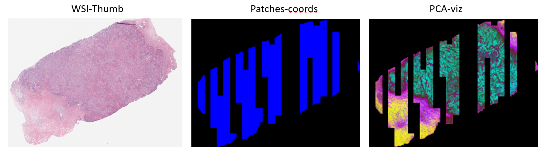
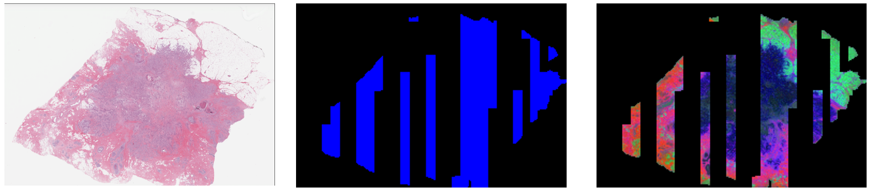
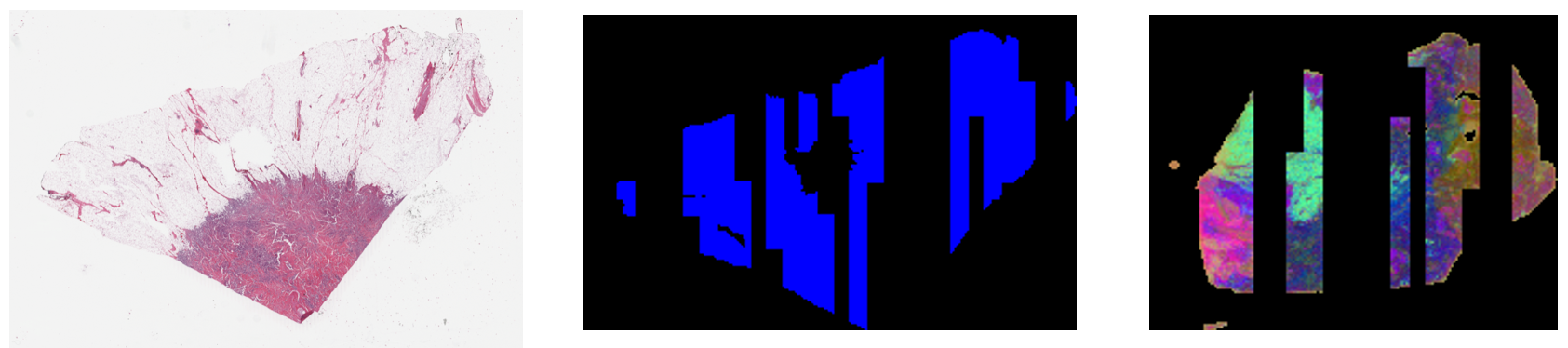
While working with TCGA-BRCA_IDC features, after some preliminary analysis I found that in some whole slide images (WSIs) viable tissue areas are discarded as in the screenshots below:
WSI GDC Link: https://portal.gdc.cancer.gov/files/d953668b-4a60-4e7c-a001-0d74e0ad6e93
WSI GDC Link: https://portal.gdc.cancer.gov/files/5f81d5d4-c4c5-4cfc-bb8e-af5bbe28db0d
WSI GDC Link: https://portal.gdc.cancer.gov/files/14a9f1fa-3ec1-4b42-bbf4-9290b0382f92
To better understand the dataset and ensure accurate downstream usage, I’d like to ask for clarification on the following points:
**Questions: **
What filtering or preprocessing criteria are applied when selecting patches?
Are you using a custom tissue mask or segmentation method to exclude background or non-informative regions?
If you could please provide some details on this that would greatly be appreciated.

It seems strange. So most background patches are filtered out but at the same time many viable areas are also discarded, right? From your results @imuhdawood , I tend to guess there could be some sampling strategies applied in the preprocessing.
Hope the authors @Richarizardd @tongding99 could provide more details about this.
Big thanks.